3D Slicer
 | |
| 原作者 | The Slicer Community |
|---|---|
| 當前版本 |
|
| 編程語言 | C++, Python |
| 操作系統 | Linux,macOS,Windows |
| 語言 | 英文 |
| 類型 | 科學視覺化軟體 |
| 許可協議 | BSD授權條款 |
| 網站 | www |
3D Slicer是開源的跨平台醫學影像視覺化和三維重建軟體。由美國國家衛生院和全球開發者社群的支援下開發。 [2]
它被廣泛用於多種醫療用途,包括自閉症、多發性硬化症、全身性紅斑狼瘡、前列腺癌、肺癌、乳腺癌、思覺失調症、骨科、生物力學、慢性阻塞性肺病、心血管疾病和神經外科。[3]
關於 3D Slicer
[編輯]3D Slicer是一個免費的開源軟體(基於BSD授權條款),用於影像分析、影像視覺化以及影像導引放射治療(Image Guided Radiotherapy,IGRT),可被用於Linux、MacOSX和windows等作業系統,它具有相當良好的可擴充性,可以透過嵌入模組的方式添加新的功能。
3D Slicer適用於查看全身各個組織的器官,相容於核磁共振造影(MRI)、電腦斷層掃描(CT)、超音波(US)以及顯微鏡下的影像。 [4]
3D Slicer支援二維多平面重建,可以用 3D 的形式將器官以不同的角度進行切割來查看不同的切面,並可結合核磁共振造影(MRI)的數據,讓醫師可以對危險程度較高的手術進行更多的事前模擬。[5]
3D Slicer的功能包括:[6]
- 處理DICOM格式的影像,並支援讀取/寫入其他種類的格式
- 支援多邊形網格、二維多平面重建以及立體渲染的視覺化
- 手動編輯影像
- 自動進行影像分割
- 擴散張量磁振造影(Diffusion Tensor Imaging,DTI)的數據分析和視覺化。
3D Slicer以BSD授權條款發佈。雖然該許可證在學術及商業用途上沒有任何限制,但是使用者有責任在遵守當地法規的情況下使用,而3D Slicer目前尚未獲得美國FDA正式批准於臨床上使用。
作業系統要求
[編輯]- Windows:Windows 10需要1903(2019年5月更新)以上的版本來支援(UTF-8)格式的文字。微軟不再支援Windows 8.1和Windows 7,所以未在這些作業系統上對3D Slicer進行過測試,但仍然可以使用。
- macOS:macOS High Sierra(10.13)或更高版本。
- Linux:Ubuntu 18.04或更高版本
- CentOS 7或更高版本。[7]
圖片集
[編輯]-
使用NVIDIA驅動程式進行硬體加速的立體渲染(此功能僅限在Windows和Linux上使用)。
-
基於感興趣區域(region of interest,ROI)的視覺化。
-
在3-Tesla磁體上獲取高分辨率數據,並使用自動跟蹤程序進行後處理。
-
腦白質影像的生成和群組分析(Group analysis)。
-
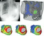
先天性心臟病患者的模型。
-
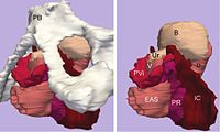
左:提肛肌的3D模型(包括恥骨和骨盆內臟)。右:沒有恥骨的相同模型。
-
從腫瘤患者影像獲得的皮質細胞碎片。
-
在手術中使用iMRI影像和3D Slicer軟體進行定位。
歷史
[編輯]3D Slicer最初是由MIT的研究生David Gering於1998年和1999年提出的,當時3D Slicer被作為布萊根婦女醫院的外科計劃實驗室與MIT人工智慧實驗室之間的碩士學位論文的一部分。[8] 四年後,3D Slicer 2於2002年發佈,可以透過FTP伺服器免費取得,當時已有數千次的下載量。2007年,3D Slicer於版本3進行了全面的改造。而3D Slicer於2009年開始下一個主要重構,它將3D Slicer採用的GUI套件從KWWidgets轉換為Qt。採用Qt的3D Slicer 4於2011年發布。[9]
用戶
[編輯]3D Slicer已被用於多種臨床研究中。在影像導引放射治療(Image Guided Radiotherapy,IGRT)的研究中,3D Slicer常被用於建立和視覺化MRI數據,這些數據在手術前和手術中均可用,並可取得用於儀器跟蹤的空間坐標。[10] 實際上,3D Slicer在影像導引放射治療中已經發揮了舉足輕重的作用,自1998年以來已有200多家出版物在文獻中引用了3D Slicer。[11]
除了從MRI影像中生成3D模型外,3D Slicer還被用於顯示從fMRI中獲得的訊息、[12] DTI(diffusion tractography) [13]和心電圖。[14] 例如,3D Slicer允許DTI影像的轉換和分析。分析的結果可以與MRI,MR血管造影和fMRI的分析結果結合。3D Slicer的其他用途包括古生物學的研究[15]和神經外科手術計劃。[16]
開發人員
[編輯]3D Slicer建立在VTK環境中,VTK是一種跨平台的圖形應用函式庫,已廣泛用於科學視覺化和ITK(Insight Segmentation and Registration Toolkit)中,ITK是一種廣泛用於開發影像分割和圖像配准的框架。在3D Slicer 4中,程式的核心是使用C ++開發而成,並且可以通過Python程式使用該API ,界面使用Qt來開發,可以使用C ++或Python進行擴充。[17]
3D Slicer支援使用輕量級的XML規範包裝任何語言的CLI程式,從中自動生成圖形使用者介面(GUI)。
對於未在3D Slicer核心程式中發佈的模組,可以使用系統自動建立和分發,以便用戶從3D Slicer中進行選擇並下載。
3D Slicer在建立過程會利用CMake自動建立必備和可選的程式庫(不包括Qt)。
參見
[編輯]參考文獻
[編輯]- ^ https://github.com/Slicer/Slicer/wiki/Release-Details#slicer-562.
- ^ 3D Slicer官方網站. [2021-02-17]. (原始內容存檔於2000-10-18).
- ^ Adriaan, Germain. 3dslicer. Brev Publishing. 2011-08-16 [2021-02-17]. ISBN 9786136666464. (原始內容存檔於2020-09-15) (英語).
- ^ 3D slicer—itread01.
- ^ 回饋社會的開源技術 3D Slicer. [2021-02-17]. (原始內容存檔於2020-08-09).
- ^ Pieper S., Lorensen B., Schroeder W., Kikinis R. The NA-MIC Kit: ITK, VTK, Pipelines, Grids and 3D Slicer as an Open Platform for the Medical Image Computing Community. Proceedings of the 3rd IEEE International Symposium on Biomedical Imaging: From Nano to Macro 2006; 1:698-701.
- ^ Getting Started—3D slicer. [2021-02-17]. (原始內容存檔於2021-04-19).
- ^ Hirayasu, Y; Shenton, ME; Salisbury, DF; Dickey, CC; Fischer, IA; Mazzoni, P; Kisler, T; Arakaki, H; Kwon, JS; Anderson, JE; Yurgelun-Todd, D; Tohen, M; McCarley, RW. Lower left temporal lobe MRI volumes in patients with first-episode schizophrenia compared with psychotic patients with first-episode affective disorder and normal subjects. The American Journal of Psychiatry. 1998, 155 (10): 1384–91. PMID 9766770. doi:10.1176/ajp.155.10.1384.
- ^ Fedorov; Beichel; Kalpathy-Cramer; Finet; Fillion-Robin; Pujol; Bauer; Jennings; Fennessy; Sonka; Buatti; Aylward; Miller; Pieper; Kikinis. 3D Slicer as an image computing platform for the Quantitative Imaging Network. Magnetic Resonance Imaging. 2012, 30 (9): 1323–41. PMC 3466397
 . PMID 22770690. doi:10.1016/j.mri.2012.05.001.
- ^ Hata, N; Piper, S; Jolesz, FA; Tempany, CM; Black, PM; Morikawa, S; Iseki, H; Hashizume, M; Kikinis, R. Application of open source image guided therapy software in MR-guided therapies. Medical Image Computing and Computer-assisted Intervention : MICCAI ... International Conference on Medical Image Computing and Computer-Assisted Intervention. 2007, 10 (Pt 1): 491–8. PMID 18051095. doi:10.1007/978-3-540-75757-3_60.
- ^ For a list of publications citing Slicer usage since 1998, visit: http://www.slicer.org/publications/pages/display/?collectionid=11 (頁面存檔備份,存於網際網路檔案館)
- ^ Archip, N; Clatz, O; Whalen, S; Kacher, D; Fedorov, A; Kot, A; Chrisochoides, N; Jolesz, F; Golby, A; Black, PM; Warfield, SK. Non-rigid alignment of pre-operative MRI, fMRI, and DT-MRI with intra-operative MRI for enhanced visualization and navigation in image-guided neurosurgery. NeuroImage. 2007, 35 (2): 609–24. PMC 3358788
 . PMID 17289403. doi:10.1016/j.neuroimage.2006.11.060.
- ^ Ziyan, U; Tuch, D; Westin, CF. Segmentation of thalamic nuclei from DTI using spectral clustering. Medical Image Computing and Computer-assisted Intervention : MICCAI ... International Conference on Medical Image Computing and Computer-Assisted Intervention. 2006, 9 (Pt 2): 807–14. PMID 17354847. doi:10.1007/11866763_99.
- ^ Verhey, JF; Nathan, NS; Rienhoff, O; Kikinis, R; Rakebrandt, F; D'ambra, MN. Finite-element-method (FEM) model generation of time-resolved 3D echocardiographic geometry data for mitral-valve volumetry. BioMedical Engineering OnLine. 2006, 5: 17. PMC 1421418
 . PMID 16512925. doi:10.1186/1475-925X-5-17.
- ^ 存档副本. [2021-02-17]. (原始內容存檔於2020-11-23).
- ^ 存档副本. [2021-02-17]. (原始內容存檔於2015-10-01).
- ^ Detection and Quantification of Small Changes in MRI Volumes. 2014: 18.


